Institutional News
JACS | De Novo Designed Long-Wavelength Fluorescence-Activating Proteins Expand the Toolkit for SWIR Bioimaging
Introduction
Fluorescence imaging has become indispensable for visualizing biological processes in cells and living organisms. However, visible light is strongly scattered by biological tissues and is often obscured by endogenous autofluorescence, limiting the performance of conventional fluorescent proteins in deep-tissue imaging.
A collaborative team led by Chunfu Xu at the National Institute of Biological Sciences, Beijing (NIBS)/Tsinghua Institute of Multidisciplinary Biomedical Research and David Baker at the University of Washington has published a study entitled “De Novo Design of Near-Infrared Fluorescence-Activating Proteins” in the Journal of the American Chemical Society. By integrating computational protein design with organic synthesis, the team created de novo proteins that bind synthetic merocyanine dyes and activate fluorescence at near-infrared (NIR) and shortwave-infrared (SWIR) wavelengths. These designed protein–dye systems provide new tools for deep-tissue imaging and establish a framework for developing future long-wavelength biosensors.
The Challenge: Opening a Longer-Wavelength Window for Deep-Tissue Imaging
Protein-based fluorescence imaging, exemplified by green fluorescent protein (GFP), has transformed modern biology. Yet visible light in the 400–700 nm range undergoes substantial scattering in biological tissues, while endogenous autofluorescence generates background that reduces imaging contrast and resolution.
The NIR region, approximately 800–1000 nm, and the SWIR region, approximately 1000–2000 nm, offer lower tissue scattering and reduced autofluorescence. In tissue phantoms, SWIR imaging has achieved penetration depths greater than 1 cm. Most existing SWIR imaging approaches, however, rely on synthetic dyes or nanomaterials and therefore lack the genetic encodability and spatiotemporal control offered by protein-based probes. Developing genetically encodable probes that operate effectively at these longer wavelengths has remained a major challenge in chemical biology and protein engineering.
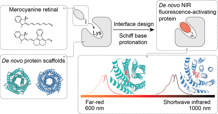
Figure 1. Design strategy for NIR and SWIR fluorescence-activating proteins
The Design Strategy: Combining Computational Protein Design with Organic Synthesis
Rather than attempting to red-shift a naturally occurring fluorescent protein, the team pursued a de novo design strategy.
The approach consisted of two complementary components:
- Fluorophore design and synthesis. The team selected and synthesized fluorogenic merocyanine retinal derivatives whose fluorescence could be activated upon conjugation to a protein and tuned toward the desired spectral range.
- Computational protein design. Using Rosetta-based design methods, the team created protein pockets that were complementary to the target dyes in both shape and chemical environment. A lysine residue was positioned at a defined site to form a covalent Schiff-base linkage with the dye. The surrounding pocket was then optimized to favor Schiff-base protonation and stabilize the resulting long-wavelength fluorescent state. A key innovation was to model the covalently linked dye–lysine conjugate as a noncanonical amino acid. This treatment enabled the dye conformation and the surrounding protein side chains to be optimized simultaneously within a unified computational framework.
Two Designed Probes Spanning the Far-Red to SWIR Regions
Using this strategy, the team developed two representative fluorescence-activating proteins.
1. MC7BP34: A Bright Far-Red Probe
MC7BP34 was designed to bind the MeroCy7 dye. Upon formation of the protein–dye complex, the excitation and emission maxima shifted to approximately 663 nm and 685 nm, respectively.
MC7BP34 was brighter than fluorescent proteins with comparable spectral properties listed in the FPbase database. In live-cell imaging experiments, the MC7BP34–MeroCy7 system enabled specific far-red labeling and showed no detectable cross-reactivity with the widely used HaloTag and SNAP-tag systems. It therefore provides an additional orthogonal channel for multicolor fluorescence imaging.
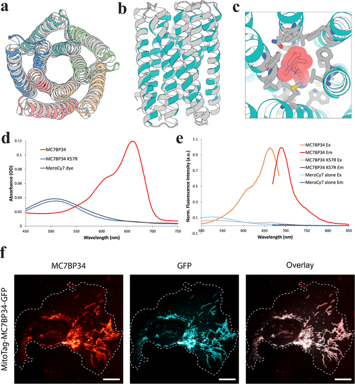
Figure 2. Design and experimental validation of the far-red fluorescence-activating protein MC7BP34
2. MC9BP81: A Long-Wavelength Probe with SWIR Emission
The team next targeted the NIR and SWIR regions by designing MC9BP81 to bind MeroCy9, a larger dye with a more extended conjugated system. The MC9BP81–MeroCy9 complex generated long-wavelength fluorescence under 892 nm excitation, with emission extending beyond 1000 nm into the SWIR region.
In deep-tissue imaging experiments in mice, the longer-wavelength MC9BP81–MeroCy9 signal was less affected by tissue autofluorescence than the signal from iRFP720 imaged under 672 nm excitation. As a result, the designed system provided improved contrast and sensitivity for identifying labeled samples through tissue.
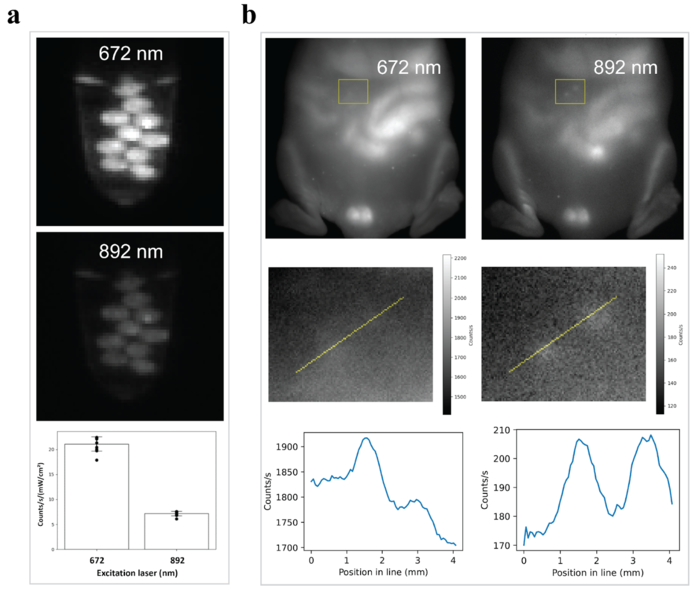
Figure 3. MC9BP81–MeroCy9 provides improved contrast and sensitivity in deep-tissue imaging compared with iRFP720
Significance and Future Directions
This study expands the toolkit available for far-red, NIR, and SWIR fluorescence imaging and demonstrates that de novo protein design can be used not only to recognize synthetic chromophores, but also to control and activate their long-wavelength photophysical properties.
Further optimization of both the dye structures and the designed binding pockets may produce NIR/SWIR probes with longer emission wavelengths, higher brightness, and improved stability under physiological conditions. Because the designed proteins are composed predominantly of α-helices, they may also be amenable to conversion into membrane-integrated sensors. Such systems could ultimately support long-wavelength imaging of electrical activity and other dynamic biological processes in deep tissues.
Authors and Funding
Chunfu Xu of the National Institute of Biological Sciences, Beijing/Tsinghua Institute of Multidisciplinary Biomedical Research and David Baker of the University of Washington are the co-corresponding authors of the study.
This work was supported by the National Key Research and Development Program of China and several international funding programs.
Article Link
https://doi.org/10.1021/jacs.5c19594