Institutional News
PNAS | Sun Shuohao’s team maps the “spatial atlas” of the vestibular ganglion, revealing the identity of gravity-sensing neurons
⭐ Highlights
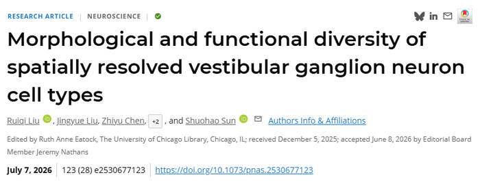
Beyond the five classical senses, the sense of balance or equilibrioception—often referred to as the “sixth sense”—maintains visual stability and body balance. However, the cellular composition of vestibular ganglion neurons (VGNs), which carry out these complex functions, has long lacked a systematic molecular definition. On July 7, 2026, the Shuohao Sun Laboratory at the National Institute of Biological Sciences, Beijing / Tsinghua Institute of Multidisciplinary Biomedical Research published a study in PNAS. Using single-cell and spatial transcriptomic approaches, the study generated the first “molecular map” and “spatial map” of VGNs, defined five functionally distinct cell types, and revealed the central role of one neuronal subtype in gravity sensing.
The vestibular system transduces motion signals into neural impulses through five end-organs in the inner ear: three semicircular canals detecting rotational acceleration, and two otolith organs sensing linear acceleration and gravity. These signals drive the vestibulo-ocular reflex, postural adjustments, and even adaptive physiological/metabolic responses (Fig. 1). Globally, approximately 15% of adults suffer from balance disorders, with prevalence exceeding 80% in individuals over 80 years old. Compared to other sensory systems, however, the molecular heterogeneity and functional specialization of vestibular neurons have remained largely a mystery.
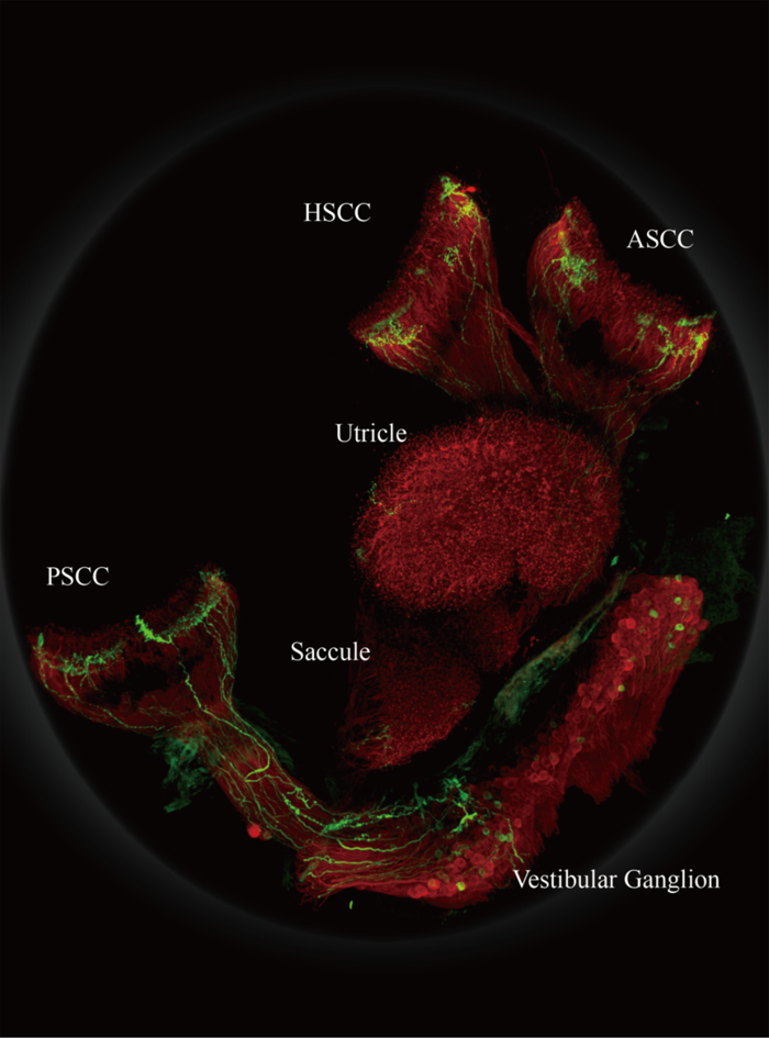
Fig. 1: Schematic of the vestibular system
The research team adopted a three-pronged strategy integrating single-cell sequencing, spatial omics, and genetic tools. They used 10x Genomics single-cell sequencing to classify cells at the molecular level; applied 10x Visium HD spatial transcriptomics to resolve the spatial coordinates of each cell type in situ; and generated CreERT2 knock-in mouse lines for each of the five cell types, enabling precise labeling and functional manipulation.
The Spatial Map of VGN Cell Types
Single-cell transcriptomics revealed five distinct VGN cell types which they named: Type A (Lypd1+/Vip+, 24.2%), Type B (F2r/Vat1l-high, 20.9%), Type C (Fxyd7+/Ddc+, 19.8%), Type D (Slc17a7+/Calb1+, 19.7%), and Type E (Pcdh17/Nkain3-high, 15.4%). Spatial transcriptomics further demonstrated that these cells are not randomly distributed but exhibit precise topological organization (Fig. 2): Type A and E cluster at the superior-inferior division boundary, Type B and C preferentially occupy the superior division, while Type D is positioned away from the boundary. Notably, in Tmie knockout mice—where hair cell mechanotransduction is abolished—the spatial positioning of Type B and D neurons shifted significantly, suggesting that sensory activity participates in the spatial arrangement of VGNs.
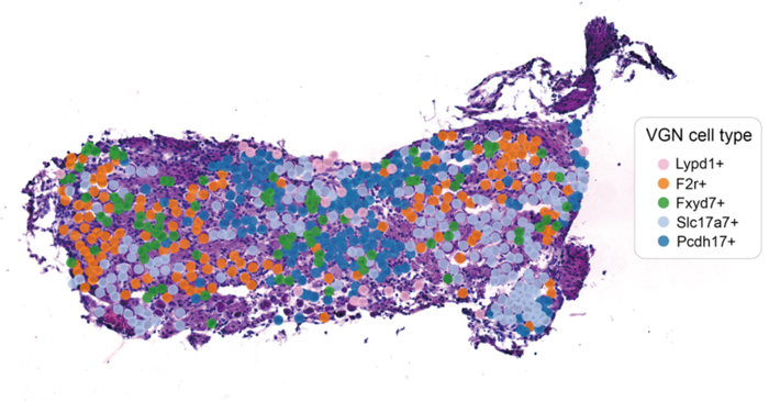
Fig. 2: Spatial atlas of VGN cell types
A Genetic Toolkit for VGN Manipulation
To precisely label and manipulate individual VGN
subtypes, the team generated a toolkit of CreERT2 knock-in mouse lines covering
all five cell types. Using this toolkit, they further mapped the peripheral and
central connectivity of these subtypes (Fig. 3). Their peripheral innervation
showed organ- and region-specific patterns: Type A/E neurons selectively
innervated the extrastriolar regions of the otolith organs, Type C neurons
specifically innervated the peripheral regions of the semicircular canals, and
Type D neurons covered the central/striolar regions of all vestibular organs.
In terms of central projections, otolith-input Type A/E neurons and
central/striolar-innervating Type D neurons densely projected to the IVN and
nucleus Y, whereas the canal-innervating Type C neurons and Type B neurons
preferentially projected to the MVNpc. This specialized “input–output”
connectivity pattern suggests that different VGN subtypes already mediate
modality-specific information segregation at the peripheral.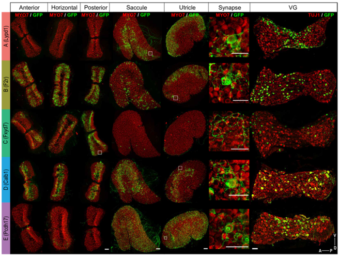
Fig. 3: Peripheral innervation patterns of the five VGN cell types
Type A VGNs: The Gravity Sensors
To directly validate their function, the team used Lypd1-DTR mice to specifically ablate Type A neurons. The results showed that postural balance and the linear vestibulo-ocular reflex (OVAR) were impaired; meanwhile, the gravitational response was completely abolished in Type A-ablated mice. Together, these findings provide the first cell-type-level evidence that Type A VGNs constitute a key cellular substrate for gravity sensing and otolith-dependent vestibular reflexes, revealing a division of labor among vestibular neuron subtypes in modality-specific information processing.
Significance and Outlook
This work provides the first systematic analysis of the heterogeneity of vestibular ganglion neurons across molecular, spatial, and functional dimensions. It challenges the traditional view that peripheral ganglion neurons are randomly distributed and establishes a complete framework linking molecular markers, spatial positioning, peripheral innervation, central projection, and behavioral function. The central role of Type A neurons in gravity sensing provides a new target for understanding the neural mechanisms underlying bone-density loss in the microgravity environment of spaceflight, and may also open potential avenues for precision treatment of vestibular dysfunction.
Team & Support
Dr. Shuohao Sun (NIBS / TIMBR) is the corresponding author. Co-first authors are Ruiqi Liu, Jingyue Liu, and Zhiyu Chen, Ph.D students in Shuohao Sun's lab. Other authors include Dr. Zhiyong Liu and Jingying Li (Ph.D student in Shuohao Sun's lab). The study was supported by the Genetic Screening Center, Imaging Facility, Transgenic Animal Center, and Bioinstrumentation Center at NIBS, and the Instrumentation Center at the Chinese Institute for Brain Research (CIBR). Dr. Ulrich Müller (Johns Hopkins University) provided valuable advice and suggestions during the course of the project. Fundings were provided by the STI-2030 Major Project and the National Key R&D Program of the Ministry of Science and Technology.
Paper link: https://www.pnas.org/doi/10.1073/pnas.2530677123